What’s the Difference Between an MRI and a CT?
Your doctor says you need a scan and immediately visions of a large machine fill your head. But do you know what you’re really in for when you need a CT or an MRI? It’s easy to get the two confused, especially if you’ve never had MRI or CT imaging done before. But from your perspective, lying on the table, there is a big difference!
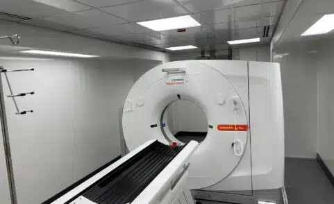
What is a CT/CAT Scan?
Wondering what the difference is between a CT and a CAT Scan. Nothing! They’re the same thing and you can use the names interchangeably. If you need a CT scan, you’re in luck, Traci says. In all her years of scanning, she’s never had a single patient who couldn’t handle a CT:
“Sometimes patients are a little bit uncomfortable in the positions we need them to hold but we’re usually able to get the scan done so quickly it’s never been a problem. Many people seem relieved that it is as easy to do as it is.”
In layman’s terms, CT is an X-ray machine that’s hooked up to a computer. You lie flat on the table and a pencil-thin beam takes cross-sectional images of your body. Traci explains that the beam rotates around your body and takes image slices of you like a loaf of bread.
CT does use radiation to capture clear images. At RAYUS, we do everything we can to make sure you are getting the lowest dose of radiation possible while still getting the highest quality images (to find out how that works, click here). The CT scan is so quick Traci says most patients’ worries about radiation exposure quickly disappear:
“It’s hard to be concerned when I explain to the patient: I’m going to ask you to hold still it’s going to be 3 minutes and I’m going to pull you right back out. I think for that reason the whole concern kind of goes away.”
CT is good for:
- Imaging bone, soft tissue and blood vessels at the same time
- Pinpointing issues with bony structures (injuries)
- Evaluating lung and chest issues (see lung scan image to the right)
- Detecting cancers
- Imaging patients with metal (no magnet)
What is an MRI Scan?
MRI stands for Magnetic Resonance Imaging. MRI uses a powerful magnetic field, radio frequency pulses and a computer to produce images of your organs, soft tissue, bone and internal structures. As a patient, you’ll lie on a table that will slide into the machine. Traditionally the space where you would go into the MRI machine was a tube or tunnel, but these days there are many options including the Wide-bore MRI, the Open MRI and the Open Upright MRI machines which give you more breathing room during your exam.
Another piece of the MR equipment is the coil. The coil acts as an antenna for the radio frequency pulses and helps the machine gather the images of a particular part of your body. The coil might be a frame that snaps over your head or shoulder, it might be placed on the table before you lie down, or it could be wrapped with Velcro around the body part that’s being scanned. To learn more about coils and see what they look like, click here. Traci explains how an MR scan of your shoulder with a coil works:
“We place your shoulder into that coil and between the radio frequency, the strong magnetic field, and the coil, which acts as an antenna, we are going to produce images of the soft tissue and the bone, with MRI.”
Even if MR is the right scan for you – you might not be right for it. If you have ever had any metal in your body, such as a pacemaker, a spinal cord stimulator, a nerve simulator, an aneurysm clip, an ear implant, or a stent, it might not be safe for you to have an MR scan. Some newer implants are safe to take into an MRI scanner, but you’ll be asked a lot of questions when you book your appointment to make sure. Orthopedic metal in the bone (like a rod in a hip or shoulder) is always going to be safe.
MRI is good for:
- Imaging organs, soft tissue an internal structures (see spine scan image to the right)
- Showing tissue difference between normal and abnormal
- Imaging without radiation
Understanding the Differences between MRI and CT
Seeing the differences laid out in a chart may help you understand how CT & MRI are unique. The shape of the machine is a major difference – CT is described as a “donut” while Traci tells her patients the MR machine is more like a “tanning bed”. CT uses radiation while MR does not. There’s also a significant difference in the length of the typical scan. Expect your MRI to take at least 30 minutes while a typical CT scan may take only 5 minutes. And while CT is great for looking at a tiny bone fracture or an organ, an MRI is better for looking at soft tissue like your brain. Remember, if you have a question about your scan, never be afraid to ask. Our technologists love to answer your questions and want you to feel relaxed and comfortable before, during and after your appointment.
What is an X-ray?
X-ray uses a small amount of radiation that passes through the body to quickly capture a single image of your anatomy to assess injury (fractures or dislocations) or disease (bone degeneration, infections or tumors). Dense objects, such as bone, block the radiation and appear white on the X-ray picture. Radiologists review the pictures and create a report with their findings to aid in diagnosis.
X-ray is good for:
- Assessing injury (see X-ray scan of the hand to the right)
Offering a low-cost, first-look exam
Guiding Your Care
Our team of specialized radiologists and clinical associates are experts in imaging. They can help guide your provider in selecting the right technology for your exam depending on your unique situation. Our goal is to provide the finest in imaging services – the right procedure, at the right time, with accurate results for each individual patient.